Understanding the Radiocarpal Joint Structure
The radiocarpal joint serves as the primary connection between your forearm and hand. This intricate articulation involves the radius bone meeting the proximal row of carpal bones. The joint functions as a condyloid synovial joint, which means it permits movement in two planes. Additionally, the joint includes the articular disc that separates the ulna from direct carpal contact. This disc acts as a cushion and stabilizer during movement.
When dealing with wrist discomfort during your travels or daily activities, having the right support equipment can make all the difference in maintaining your mobility and comfort. If you’re experiencing mild wrist strain from carrying luggage or working on your laptop during long flights, consider using a wrist compression strap that provides adjustable support without limiting your range of motion too severely. For travelers who need more substantial support throughout their journey, an adjustable wrist brace can offer reversible protection that works for both hands and helps prevent tendonitis flare-ups during extended periods of activity. Many digital nomads and frequent travelers swear by copper compression sleeves because they’re comfortable enough to wear throughout the day while providing gentle support for arthritis and carpal tunnel symptoms. If you’re working remotely from various locations and spending hours typing travel blog posts or editing photos, investing in an ergonomic keyboard wrist rest with memory foam can significantly reduce strain and help prevent repetitive stress injuries from developing over time. To maintain wrist strength and prevent injuries before they occur, incorporating a grip strengthener kit into your daily routine can help build the forearm muscles that support proper wrist mechanics during all your adventures. For more targeted strengthening exercises that you can do in your hotel room or Airbnb, a wrist rotation exerciser allows you to perform specific movements that improve grip strength and wrist stability without requiring much space in your luggage. When existing wrist pain is already affecting your travel experience, an orthopedic wrist splint with copper infusion can provide the rigid support necessary for healing while you continue exploring new destinations with modified activities. After particularly strenuous days of hiking, climbing, or carrying heavy camera equipment, applying a therapeutic ice pack wrap can reduce inflammation and provide soothing relief that helps you recover faster for the next day’s adventures. If you’re editing photos or managing your travel social media accounts from cafes and coworking spaces around the world, pairing your laptop with an ergonomic mouse pad featuring gel wrist support can prevent discomfort during those marathon editing sessions that every travel blogger knows too well. Finally, for rehabilitation after a wrist injury or for preventing tennis elbow from activities like paddleboarding or rock climbing during your travels, incorporating a flex therapy bar into your recovery routine can help restore strength and flexibility so you can get back to fully enjoying all the physical activities that make travel so rewarding and memorable.
The joint capsule surrounding this area contains synovial fluid that lubricates the surfaces. Furthermore, several ligaments reinforce the joint’s stability. These include the palmar radiocarpal ligaments and the dorsal radiocarpal ligaments. Meanwhile, the radial and ulnar collateral ligaments provide lateral support. This complex network of soft tissues prevents excessive movement while maintaining flexibility. Source ## Range of Motion Capabilities
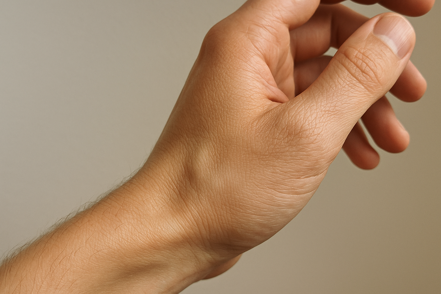
The wrist joint enables four primary movement directions that are essential for daily activities. Flexion allows you to bend your hand toward your palm. Conversely, extension lets you bend your hand backward. Additionally, the joint permits radial deviation, moving your hand toward your thumb side. Similarly, ulnar deviation moves your hand toward your pinky finger side.
These movements combine to create circumduction, a circular motion of the hand. For example, when you stir a pot or wave goodbye, you’re using circumduction. The typical range of flexion reaches approximately 80 degrees. In contrast, extension usually achieves about 70 degrees. Moreover, radial deviation allows roughly 20 degrees of movement. Meanwhile, ulnar deviation permits approximately 30 degrees of motion.
Clinical Significance and Common Pathologies
Healthcare professionals must thoroughly understand wrist joint anatomy for accurate diagnosis. Carpal tunnel syndrome represents one of the most prevalent conditions affecting this region. This disorder occurs when the median nerve becomes compressed within the carpal tunnel. Furthermore, wrist sprains frequently result from falls onto an outstretched hand.
Fractures of the distal radius are particularly common among older adults. These injuries often require immediate medical attention and proper immobilization. Additionally, arthritis can develop in the radiocarpal joint over time. Rheumatoid arthritis and osteoarthritis both affect this joint’s function. Therefore, early recognition of symptoms leads to better treatment outcomes. Consequently, understanding normal wrist anatomy helps identify abnormal conditions quickly.
Articular Surfaces
Anatomical Structure of the Radiocarpal Joint
The radiocarpal joint represents a critical articulation point in the wrist. This joint forms where the forearm meets the hand. The distal radius creates the primary bony foundation for this connection. Meanwhile, three carpal bones contribute to the joint’s distal component.
The radius bone features a distinctly concave articular surface at its distal end. This depression is specifically shaped to accommodate the wrist bones. Furthermore, this concave design allows for smooth gliding movements. The surface is divided into two distinct facets. Additionally, these facets provide separate contact points for different carpal bones.
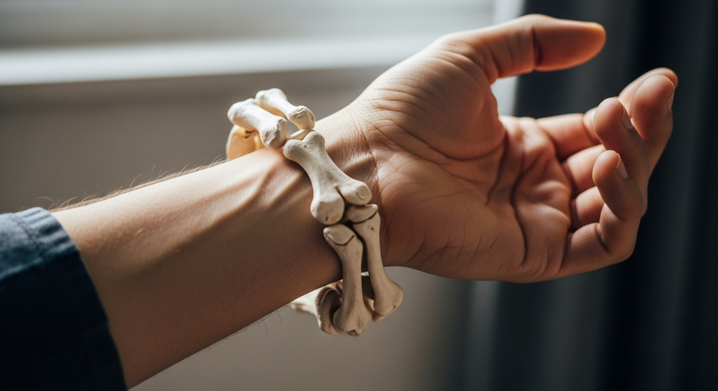
The Carpal Bone Contributors
Three specific carpal bones form the proximal row that articulates with the radius. The scaphoid bone sits on the thumb side of the wrist. Moreover, it occupies the largest contact area with the radius. The lunate bone positions itself in the middle of the proximal row. Consequently, it serves as the central pivot point for wrist motion. The triquetrum bone completes the proximal row on the pinky side.
These three bones present convex articular surfaces that fit into the radius. For example, imagine a ball resting in a shallow socket. This convex-concave relationship creates what anatomists call an ellipsoid joint. Therefore, the wrist can move in multiple directions simultaneously.
Movement Capabilities and Range
The radiocarpal joint’s unique design enables impressive wrist flexibility. The joint allows flexion, bringing the palm toward the forearm. Additionally, extension moves the hand backward. Radial deviation tilts the hand toward the thumb side. In contrast, ulnar deviation angles the hand toward the pinky finger.
However, the joint’s capabilities extend beyond simple planar movements. Circumduction combines all these motions into circular patterns. As a result, the hand can trace cone-shaped movements through space. This versatility proves essential for daily activities like writing and typing.
The Ulna’s Indirect Role
The ulna bone runs parallel to the radius in the forearm. However, it does not directly contact the carpal bones. Instead, a specialized structure bridges this gap. The triangular fibrocartilage complex (TFCC) fills the space between ulna and carpals.
The TFCC consists of fibrocartilage tissue and supporting ligaments. Moreover, it acts as a cushioning pad during wrist movements. This structure distributes forces across the ulnar side of the wrist. Consequently, it prevents excessive stress on any single bone. Furthermore, the TFCC enhances overall joint stability during rotational movements.
The complex also allows the radius and ulna to rotate around each other. Therefore, forearm rotation becomes possible without disrupting wrist integrity. Additionally, the TFCC contributes to load-bearing during gripping activities. Source
Joint Capsule and Ligaments
The Protective Fibrous Capsule
The wrist’s fibrous capsule serves as a protective sleeve around the joint. This dense connective tissue structure wraps completely around the radiocarpal articulation. Furthermore, it extends distally to encompass portions of the intercarpal joints. The capsule consists of tightly woven collagen fibers that resist stretching. These fibers provide essential mechanical support during daily activities. Additionally, the capsule attaches firmly to the radius, ulna, and proximal carpal bones. This attachment pattern creates a secure enclosure for the joint space.
The capsule’s thickness varies considerably across different regions of the wrist. Moreover, certain areas contain specialized thickenings that form distinct ligamentous bands. These reinforced zones correspond to areas experiencing the greatest mechanical stress. The capsule also produces synovial fluid through its inner lining. This fluid lubricates the joint surfaces and nourishes the cartilage.
Dorsal and Palmar Radiocarpal Ligaments
The dorsal radiocarpal ligament originates from the posterior aspect of the distal radius. It fans out obliquely to insert on the triquetrum and lunate bones. This ligament primarily restricts excessive palmar flexion of the hand. For example, when you bend your wrist backward, this ligament becomes taut. Consequently, it prevents hyperextension that could damage delicate joint structures.
In contrast, the palmar radiocarpal ligaments are considerably stronger and more complex. These ligaments originate from the palmar surface of the radius and ulna. They consist of multiple distinct bands with different attachment points. The radioscaphocapitate ligament represents one important component of this system. Meanwhile, the radiolunate and radioscapholunate ligaments provide additional support. Source These structures work synergistically to control dorsiflexion and maintain carpal alignment.
Maintaining Carpal Bone Alignment
The radiocarpal ligaments create a sophisticated stabilization system for the eight carpal bones. These small bones must maintain precise spatial relationships during movement. Therefore, the ligaments act like architectural cables in a suspension bridge. They distribute forces evenly across the joint surfaces. Additionally, they guide the carpal bones through their normal range of motion.
The ligaments prevent abnormal translation or shifting of individual carpal bones. For instance, during forceful gripping activities, compression forces increase dramatically. However, the ligamentous network resists these destabilizing forces effectively. Moreover, proprioceptive nerve endings within the ligaments provide sensory feedback. This feedback helps the brain monitor wrist position continuously. As a result, the neuromuscular system can make rapid adjustments to maintain stability.
Limiting Excessive Movements
Each ligament has a specific range of motion it permits before becoming taut. When you move your wrist through flexion, extension, or deviation, different ligaments engage sequentially. This sequential engagement creates a smooth, controlled motion pattern. Furthermore, the ligaments prevent movements that exceed safe anatomical limits.
Excessive radial deviation, for example, stretches the ulnar-sided ligaments maximally. Conversely, extreme ulnar deviation tensions the radial-sided structures. The ligaments essentially define the boundaries of safe wrist motion. Additionally, they absorb and dissipate energy during sudden impacts or falls. This protective mechanism reduces the risk of bone fractures and cartilage damage.
Collateral Ligament Contributions
The ulnar collateral ligament extends from the ulnar styloid process distally. It inserts primarily on the triquetrum and pisiform bones. This ligament provides critical resistance against radial deviation forces. Moreover, it stabilizes the ulnar aspect of the wrist during power grip activities. Many occupational tasks, such as hammering or twisting motions, stress this ligament significantly.
The radial collateral ligament originates from the radial styloid process. It courses obliquely to attach on the scaphoid bone and trapezium. This structure prevents excessive ulnar deviation of the hand. Furthermore, it maintains the proper relationship between the radius and proximal carpal row. Athletes who participate in racquet sports particularly depend on this ligament’s integrity.
Preventing Dislocation and Lateral Instability
The collateral ligaments function as lateral stabilizers that resist sideways displacement. Without these structures, the carpus could subluxate or dislocate medially or laterally. For example, a fall on an outstretched hand generates substantial lateral shearing forces. However, intact collateral ligaments typically prevent complete joint disruption.
These ligaments also work in coordination with the radiocarpal ligaments. Together, they create a three-dimensional stability matrix around the wrist joint. Additionally, the collateral ligaments help maintain even pressure distribution across articular surfaces. This balanced loading pattern minimizes localized cartilage wear over time. Consequently, the ligamentous system contributes significantly to long-term joint health and function.
Blood Supply and Innervation
Vascular Anatomy of the Wrist
The vascular network surrounding the carpal joint is remarkably complex. Primarily, the radial and ulnar arteries provide the essential blood flow required for tissue health. These two major vessels do not function in isolation. Instead, they interconnect to form intricate structures known as the superficial and deep palmar arches. This redundancy creates a safety net for the hand. Consequently, blood flow remains consistent even when the wrist twists or flexes.
However, the distribution of blood is not uniform across all carpal bones. For instance, the scaphoid bone relies on a retrograde blood supply. This means blood flows backward from the distal end to the proximal end. As a result, fractures in this area carry a high risk of poor healing. Therefore, maintaining healthy circulation is critical for preserving long-term joint integrity.
Neurological Control and Sensation
Beyond circulation, the carpal joint serves as a major thoroughfare for the nervous system. Three distinct nerves coordinate to provide both motor control and sensory feedback. Specifically, the median, ulnar, and radial nerves work in unison to enable dexterity. Without this precise innervation, simple tasks like gripping a pen would be impossible.
Each nerve manages a specific zone of the hand:
- Median Nerve: This nerve travels through the carpal tunnel. Crucially, it supplies sensation to the thumb, index, and middle fingers.
- Ulnar Nerve: Often called the “funny bone” nerve, it controls fine muscle movements. Additionally, it provides feeling to the little finger and half of the ring finger.
- Radial Nerve: This structure is responsible for wrist extension. Furthermore, it supplies sensation to the back of the hand.
Functional Implications
The synergy between these vascular and neural systems facilitates complex hand function. The nerves transmit signals for movement, while the arteries fuel the muscles executing those commands. Consequently, any impingement or trauma can cause significant disability.
For example, compression of the median nerve leads to Carpal Tunnel Syndrome. Symptoms often include numbness, tingling, or weakness. Similarly, vascular damage can lead to tissue necrosis in the carpal bones. Thus, protecting these anatomical structures is vital for overall upper limb health.
Movements of the Wrist Joint
Understanding Wrist Mechanics
The carpal joint acts as a complex bridge between the forearm and the hand. It is not simply a hinge that moves back and forth. Instead, it offers a dynamic range of motion across multiple planes. Consequently, this flexibility allows humans to interact with the world effectively. The joint structure is composed of eight small carpal bones. These bones glide against each other to facilitate smooth articulation.
Furthermore, the wrist relies on a sophisticated network of ligaments for stability. Without this support, the joint would be too loose for heavy tasks. Therefore, the carpal joint balances mobility with necessary structural strength. This unique combination is vital for human dexterity.
Primary Movements: Flexion and Extension
Flexion serves as a primary movement for the wrist complex. Specifically, this action decreases the angle between the palm and the inner forearm. Imagine curling your hand inward to check a watch. Source Furthermore, powerful flexor muscles in the forearm drive this motion. Without flexion, gripping objects would be nearly impossible.
Extension functions as the direct counterpart to flexion. During this movement, the back of the hand moves closer to the forearm. For example, consider the motion used when pushing open a heavy door. Additionally, extension stabilizes the wrist during strong gripping actions. This stability prevents the wrist from collapsing under pressure.
Key differences include:
- Flexion: Bends the palm inward (e.g., throwing a basketball).
- Extension: Pulls the knuckles back (e.g., doing a push-up).
Lateral Movements: Abduction and Adduction
Radial deviation, technically known as abduction, involves moving the wrist toward the thumb side. This movement occurs along the frontal plane. Structurally, the radius bone limits this motion slightly. Nevertheless, it remains crucial for precise tasks. For instance, you use this motion when waving goodbye or swinging a hammer.
Conversely, ulnar deviation (adduction) shifts the hand toward the pinky finger. This range of motion is typically greater than radial deviation. The anatomy of the ulna allows for this wider sweep. Therefore, activities like chopping vegetables rely heavily on this specific articulation. It provides the necessary angle for fluid hand positioning.
Functional Importance in Daily Life
All these movements work together seamlessly. Rarely do they happen in total isolation during daily life. As a result, the carpal joint enables complex actions like typing or throwing a ball. Even simple tasks, such as pouring water, require a blend of these motions.
Moreover, maintaining flexibility here is vital for overall hand health. Stiffness in the carpal joint can severely limit independence. Ultimately, proper wrist function defines our manual dexterity. It transforms simple muscle contractions into refined, useful tools.
Common Clinical Correlations
The wrist is prone to various injuries, with Colles fracture being one of the most common. Source This fracture occurs at the distal radius and is often caused by falling onto an outstretched hand. Treatment typically involves immobilization or surgical intervention, depending on the severity .
Understanding Wrist Instability
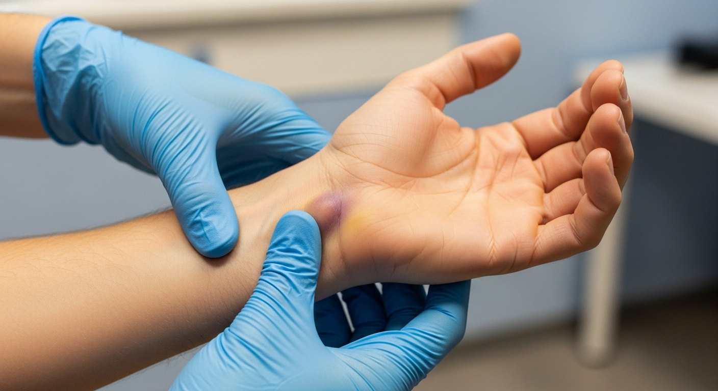
Ligament damage represents one of the most common causes of wrist dysfunction. These injuries typically occur during falls, sports activities, or repetitive strain. When ligaments tear or stretch beyond their capacity, the bones lose proper support. Consequently, the wrist becomes unstable and painful during movement.
The symptoms of ligament-related instability vary significantly among patients. Some individuals experience sharp, shooting pains during specific movements. Others notice a dull, persistent ache throughout the day. Additionally, many patients report a clicking or popping sensation when rotating their wrist. Furthermore, swelling often develops around the affected joint area.
Range of Motion Limitations
Movement restrictions significantly impact daily activities and quality of life. Simple tasks like opening jars or turning doorknobs become challenging. Moreover, gripping objects firmly may prove difficult or impossible. The wrist may feel weak when bearing weight during push-ups or similar exercises.
Chronic instability can lead to progressive joint deterioration if left untreated. For example, cartilage damage may develop over time. The bones may shift from their normal positions gradually. As a result, arthritis can develop in the carpal joints prematurely.
Treatment Approaches for Recovery
Physical therapy serves as the first-line treatment for mild to moderate instability. Therapists design customized exercise programs to strengthen supporting muscles. These exercises focus on improving grip strength and wrist stability. Additionally, therapists may use manual techniques to restore proper joint mechanics.
Treatment protocols typically include several key components:
- Strengthening exercises targeting forearm and hand muscles
- Flexibility training to restore normal range of motion
- Proprioceptive drills improving joint position awareness
- Functional activities simulating real-world tasks
- Modalities like ultrasound or electrical stimulation for pain management
However, severe ligament tears often require surgical intervention. Surgeons may perform ligament reconstruction using tissue grafts. Therefore, they can stabilize the joint and prevent further damage.
Diagnostic Importance of Anatomical Knowledge
Precise diagnosis depends heavily on understanding carpal joint structure and function. Clinicians must recognize how eight small carpal bones interact with each other. These bones form complex articulations that allow multidirectional wrist movement. Meanwhile, numerous ligaments connect these bones in specific patterns.
Medical professionals use this anatomical knowledge during physical examinations. For instance, they perform specific stress tests targeting individual ligaments. These tests reveal which structures have sustained damage. Furthermore, imaging studies like MRI scans confirm clinical findings.
Mechanics and Treatment Planning
Biomechanical principles guide effective treatment strategies for wrist instability. Clinicians analyze how forces transfer through the carpal bones during movement. They identify which ligaments provide primary versus secondary stability. Consequently, this understanding helps determine whether conservative or surgical treatment is appropriate.
Treatment plans must address the specific mechanical dysfunction present. For example, scapholunate ligament injuries require different approaches than lunotriquetral tears. Moreover, the patient’s activity level and occupation influence treatment decisions significantly.
Conclusion
The Complexity of Carpal Mechanics
The human wrist functions as a highly sophisticated mechanical system. It is far more than a simple hinge. Specifically, the interaction between the forearm and hand bones facilitates intricate maneuvers. These movements are necessary for interacting with our environment. Consequently, humans can perform delicate tasks with extreme precision. For instance, writing requires subtle micro-adjustments of the carpal bones.
Key movements include:
- Flexion: Bending the palm downward.
- Extension: Raising the back of the hand.
- Radial/Ulnar Deviation: Moving the hand sideways.
Without this mobility, basic actions would become impossible. Therefore, the joint’s design prioritizes both flexibility and strength.
Anatomy and Structural Integrity
Several anatomical layers ensure the joint remains stable under stress. The articular surfaces must remain smooth and lubricated. This allows the bones to glide without friction. Furthermore, a complex network of ligaments binds these bones together. These ligaments act as strong biological cables. Source As a result, they limit excessive motion during heavy lifting or impact.
Crucial anatomical components:
- Carpal Bones: Two rows of small bones forming the wrist base.
- Radius and Ulna: The forearm bones providing support.
- Articular Cartilage: The protective coating on bone ends.
However, this complexity creates vulnerability. If one ligament fails, the entire structure may destabilize. Thus, maintaining ligament health is essential for long-term function.
Vital Supply Lines: Blood and Nerves
Proper joint function relies heavily on vascular and neurological health. Blood vessels nourish the living cartilage and bone. The radial and ulnar arteries are the primary sources of this fuel. Additionally, the nervous system controls every specific movement. Nerves transmit instant signals from the brain to the hand.
Moreover, these nerves provide critical sensory feedback. They tell us if an object is hot, cold, or sharp. In contrast, poor circulation can lead to tissue death. Similarly, nerve damage often results in numbness or weakness. Consequently, protecting these supply lines is a priority in any wrist surgery.
Clinical Applications and Future Tech
Medical professionals rely on this detailed anatomical knowledge daily. It directly guides the treatment of common ailments. For example, treating Carpal Tunnel Syndrome requires understanding nerve pathways. Accurate diagnosis leads to better surgical plans and physical therapy. Meanwhile, researchers are constantly developing new techniques.
Recent advancements include:
- Arthroscopy: Minimally invasive surgical procedures.
- 3D Printing: Custom implants for severe fractures.
- Biologics: Injections that promote natural tissue healing.
Furthermore, modern imaging helps significantly. MRI scans now reveal minute details of soft tissue damage. As a result, surgeons can operate with greater accuracy than ever before. This leads to faster recovery times for patients. Ultimately, technology continues to restore function and improve quality of life. Source
When dealing with wrist discomfort during your travels or daily activities, having the right support equipment can make all the difference in maintaining your mobility and comfort. If you’re experiencing mild wrist strain from carrying luggage or working on your laptop during long flights, consider using a wrist compression strap that provides adjustable support without limiting your range of motion too severely. For travelers who need more substantial support throughout their journey, an adjustable wrist brace can offer reversible protection that works for both hands and helps prevent tendonitis flare-ups during extended periods of activity. Many digital nomads and frequent travelers swear by copper compression sleeves because they’re comfortable enough to wear throughout the day while providing gentle support for arthritis and carpal tunnel symptoms. If you’re working remotely from various locations and spending hours typing travel blog posts or editing photos, investing in an ergonomic keyboard wrist rest with memory foam can significantly reduce strain and help prevent repetitive stress injuries from developing over time. To maintain wrist strength and prevent injuries before they occur, incorporating a grip strengthener kit into your daily routine can help build the forearm muscles that support proper wrist mechanics during all your adventures. For more targeted strengthening exercises that you can do in your hotel room or Airbnb, a wrist rotation exerciser allows you to perform specific movements that improve grip strength and wrist stability without requiring much space in your luggage. When existing wrist pain is already affecting your travel experience, an orthopedic wrist splint with copper infusion can provide the rigid support necessary for healing while you continue exploring new destinations with modified activities. After particularly strenuous days of hiking, climbing, or carrying heavy camera equipment, applying a therapeutic ice pack wrap can reduce inflammation and provide soothing relief that helps you recover faster for the next day’s adventures. If you’re editing photos or managing your travel social media accounts from cafes and coworking spaces around the world, pairing your laptop with an ergonomic mouse pad featuring gel wrist support can prevent discomfort during those marathon editing sessions that every travel blogger knows too well. Finally, for rehabilitation after a wrist injury or for preventing tennis elbow from activities like paddleboarding or rock climbing during your travels, incorporating a flex therapy bar into your recovery routine can help restore strength and flexibility so you can get back to fully enjoying all the physical activities that make travel so rewarding and memorable.
*As an Amazon Associate, I earn from qualifying purchases.