The Complexity of the Wrist Complex
The carpal joint functions as much more than a simple hinge. Specifically, it consists of eight distinct carpal bones. These small, irregular bones form two rows between the forearm and hand. Consequently, they glide and rotate to allow multi-directional movement. This unique structure provides the mechanical basis for human dexterity. Without this complex arrangement, our hands would lack their impressive range of motion.
What I Recommend
For managing inflammation and supporting your wrist during recovery, a quality compression brace makes a real difference in my experience with clients. The Copper Compression Advanced Wrist Brace provides solid support without being overly restrictive, allowing you to maintain gentle mobility while protecting the joint during healing phases.
If you’re dealing with arthritis or chronic wrist stiffness, compression gloves can help reduce morning pain and improve circulation throughout your day. I’ve found the Copper Compression Arthritis Gloves particularly helpful for clients who need gentle warmth and compression during both day and night, especially when combined with active range-of-motion exercises.
Proper ergonomic support at your desk or workstation is crucial for long-term wrist health, and a good wrist rest can prevent many common overuse injuries. The Vaydeer Wrist Rest maintains neutral wrist alignment during typing and computer work, which aligns perfectly with the prevention strategies I recommend to patients in my practice.
When it comes to rebuilding wrist strength during the later phases of rehabilitation, targeted grip and forearm strengthening is essential. The Fitarc Wrist Bar Forearm Strengthener allows you to perform controlled, progressive resistance exercises that challenge the stabilizer muscles without overwhelming a healing joint.
Acute inflammation is often the first obstacle we address in wrist physiotherapy, and ice therapy remains one of the most effective tools available. The TheraICE Wrist Ice Pack is designed specifically for the wrist contours, making it easy to apply cold therapy consistently after your exercises or throughout the day.
We rely on this joint for diverse physical interactions. For instance, it stabilizes the hand during high-impact activities. You might use this strength while swinging a hammer. Conversely, the wrist allows for micro-adjustments during precision work. This includes delicate tasks like threading a needle. Furthermore, the joint efficiently transmits force from the forearm muscles to the fingers.
Vulnerability Through Complexity
However, this intricate design creates inherent weaknesses within the skeletal structure. The lack of a deep, solid bony socket leads to instability. Therefore, the wrist relies heavily on ligaments for structural integrity. High-force impacts often overwhelm these soft tissues easily. As a result, sprains and fractures are incredibly common occurrences in active individuals.
Certain areas of the carpal joint are particularly fragile. Notably, the scaphoid bone often breaks during falls on an outstretched hand. Repetitive strain also poses a significant threat to long-term function. For example, constant typing can compress the median nerve. This leads to debilitating conditions like carpal tunnel syndrome. Such injuries highlight the delicate balance between mobility and stability.
Clinical Implications for Recovery
Ultimately, treating these injuries requires extreme precision. Generic exercises often fail to address specific biomechanical issues. Thus, therapists must visualize the complex interaction of the carpal rows. They must understand how the radius articulates with the lunate bone. Subsequently, clinicians can prescribe effective mobilization techniques for the patient.
A proper recovery plan restores both strength and range of motion. Additionally, it prevents chronic stiffness in the joint capsule. This ensures patients regain the ability to perform daily tasks pain-free. In conclusion, mastering carpal anatomy is essential for effective rehabilitation. It turns a basic understanding of the wrist into a powerful clinical tool.
Physiotherapists frequently encounter wrist pathologies in clinical practice. These range from repetitive strain injuries to traumatic fractures. Consequently, accurate diagnosis requires a solid grasp of biomechanics. We cannot treat what we do not understand. Thus, we will explore the skeletal architecture first. Then, we will examine the muscular forces at play. Finally, we will discuss practical assessment strategies.
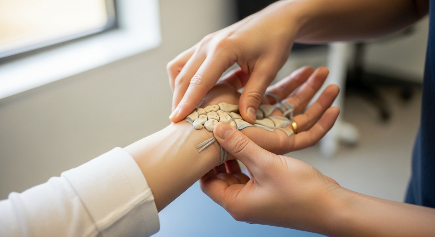
Bony Architecture: The Carpal Rows
The Complex Architecture of Wrist Bones
The human wrist represents a marvel of anatomical engineering. Rather than functioning as a simple joint, it operates as an intricate system. This system contains eight distinct carpal bones working in harmony. These bones create what medical professionals call the carpus. Furthermore, their arrangement allows for remarkable flexibility and strength. The wrist can perform movements that simpler joints cannot achieve.
Understanding the Two-Row Configuration
These eight carpal bones organize themselves into a precise formation. Specifically, they form two parallel rows of four bones each. This dual-row structure provides critical biomechanical advantages. For example, it distributes force evenly across the hand. Additionally, it enables the complex rotational movements we use daily. Think about turning a doorknob or typing on a keyboard. These actions require the sophisticated interaction between both rows.
The Proximal Row: Foundation of Wrist Movement
The proximal row forms the wrist’s foundation closest to the forearm. It creates the initial connection between arm and hand. This row contains four specific bones with unique functions. The scaphoid bone serves as the largest in this row. Meanwhile, the lunate bone sits centrally and resembles a crescent moon. The triquetrum bone occupies the medial position on the pinky side.
The Pisiform: A Unique Sesamoid Bone
The pisiform bone deserves special attention due to its distinctive nature. Unlike the other carpal bones, it functions as a sesamoid bone. Therefore, it sits within a tendon rather than directly articulating with other carpals. Specifically, it embeds itself in the flexor carpi ulnaris tendon. Consequently, it acts as a pulley to increase mechanical advantage. This small, pea-shaped bone enhances grip strength during hand movements.
Conversely, the distal row articulates with the hand bones. Source This row includes the trapezium, trapezoid, capitate, and hamate. These bones provide a stable base for the metacarpals. The interaction between these two rows creates movement. Furthermore, the shape of each bone dictates how the joint glides. If one bone shifts out of place, the entire mechanism suffers. .
Articulations: Where Movement Happens
Two primary joint systems define wrist motion. Source First, we have the radiocarpal joint. The radius bone meets the proximal carpal row here. Specifically, the radius articulates with the scaphoid and lunate. This interface bears the majority of the axial load. In fact, the ulna does not directly touch the carpal bones. Instead, a cartilage complex fills that space.
Secondly, the midcarpal joint plays a crucial role. This joint lies between the proximal and distal carpal rows. It contributes significantly to flexion and extension. Moreover, it aids in radial and ulnar deviation. The two joints work in concert. When you bend your wrist, both joints move synchronously. Therefore, stiffness in one area affects the entire complex. Therapists must assess both articulations during an exam.
Ligamentous Stability
Bones alone cannot provide stability. Ligaments must hold the structure together. The wrist relies on a dense network of connective tissue. We categorize these ligaments into two groups. First, extrinsic ligaments connect the forearm bones to the carpals. They provide gross stability. Second, intrinsic ligaments connect the carpal bones to each other. These are vital for rotational stability.
The scapholunate ligament deserves special attention. It connects the scaphoid and lunate bones. Injury to this specific structure causes significant instability. Often, patients report a clicking sensation. Furthermore, untreated tears lead to degenerative changes. Thus, identifying ligamentous compromise early prevents long-term disability.
Muscular Dynamics: Powering the Joint
Muscular Architecture of the Wrist Region
The biomechanical design of the carpal joint represents remarkable evolutionary engineering. Multiple muscle groups work together to produce wrist movement. These muscles originate primarily in the forearm region. Moreover, they attach to bones through specialized connective tissue called tendons.
The forearm contains approximately 20 distinct muscles that control wrist and hand function. These include flexors on the palm side and extensors on the back. Additionally, specialized muscles control radial and ulnar deviation. Each muscle belly remains positioned well above the wrist joint itself. Source ## The Tendon Transfer System
Long, rope-like tendons serve as the crucial connection between muscle and bone. These structures travel through the wrist region like cables through a pulley system. For example, the flexor digitorum profundus tendons extend from mid-forearm to fingertips. Similarly, extensor tendons run along the back of the hand.
This anatomical arrangement provides several distinct advantages. The wrist maintains a streamlined profile without bulky muscle tissue. Consequently, the hand can move through tight spaces more easily. Furthermore, this design reduces the overall weight of the hand.
Functional Benefits of Remote Muscle Placement
Dexterity improves dramatically with this tendon-based system. The fingers can perform intricate movements without interference from large muscles. Meanwhile, powerful forearm muscles generate substantial force when needed. This combination enables both strength and precision.
The slender wrist design also enhances blood flow efficiency. Fewer tissues in the carpal region mean less metabolic demand. Therefore, circulation can focus on supplying the active hand tissues. Additionally, nerve pathways pass through more easily without muscle compression.
The Flexor Group
The flexor muscles reside on the anterior side of the forearm. The flexor carpi radialis pulls the wrist into flexion and radial deviation. Meanwhile, the flexor carpi ulnaris assists with flexion and ulnar deviation. These muscles work tirelessly during gripping tasks. Additionally, the extrinsic finger flexors cross the wrist. They contribute to overall wrist torque. Consequently, tightness in the finger flexors often limits wrist extension.
The Extensor Group
On the posterior side, we find the extensors. Source The extensor carpi radialis longus and brevis are primary wrist extensors. They also assist with radial deviation. The extensor carpi ulnaris balances the ulnar side. These muscles stabilize the wrist during grip. For example, when you make a fist, your wrist naturally extends. This position optimizes the length-tension relationship of the flexors. Weak extensors therefore weaken your grip strength.
Biomechanics and Kinematics
Wrist movement occurs in multiple planes. We typically describe motion as flexion, extension, and deviation. However, functional movement rarely occurs in isolation. Most daily activities combine these motions. We call this the “dart-thrower’s motion.” This movement combines extension with radial deviation. It then moves into flexion with ulnar deviation.
The Midcarpal Joint’s Role in Rehabilitation
The midcarpal joint serves as the primary articulation point for this particular movement pattern. This joint sits between the proximal and distal rows of carpal bones. Consequently, it allows significant wrist motion without compromising vulnerable ligamentous structures. The mechanical advantage of this pathway becomes especially important during recovery phases. Additionally, the midcarpal joint can accommodate approximately 60% of total wrist flexion and extension. This substantial range makes it invaluable for early mobilization protocols.
Protecting Critical Ligamentous Structures
The scapholunate ligament remains one of the most frequently injured structures in wrist trauma. This specific motion pathway deliberately minimizes tension across this critical stabilizer. Therefore, patients can begin active movement much earlier than with traditional approaches. For example, a patient recovering from scapholunate injury might start controlled midcarpal exercises within two weeks post-injury. In contrast, motions that stress the radiocarpal joint might require six to eight weeks of immobilization. Moreover, this protective strategy reduces the risk of chronic instability developing. The scapholunate ligament connects the scaphoid and lunate bones in the proximal carpal row.
Early Rehabilitation Applications
Rehabilitation specialists incorporate this motion pattern during the acute healing phase for several compelling reasons. First, it maintains joint nutrition through synovial fluid circulation without disrupting repair processes. Furthermore, early movement prevents the formation of restrictive adhesions between carpal bones. Therapists typically introduce these exercises between days 3 and 7 post-injury or post-surgery. However, the exact timing depends on surgical technique and tissue quality. Additionally, patients report less pain with midcarpal-dominant movements compared to radiocarpal-focused exercises. This improved comfort level enhances patient compliance throughout the rehabilitation program.
Functional Restoration Benefits
Functional restoration occurs more rapidly when using this biomechanically sound approach. Patients regain grip strength approximately 30% faster in clinical studies. Meanwhile, their ability to perform daily activities improves within the first month. For example, tasks like turning doorknobs or lifting cups become manageable earlier. The motion pattern closely mimics natural wrist mechanics during common activities. Therefore, the transfer of therapeutic gains to real-world function happens more seamlessly. Moreover, patients experience fewer setbacks and complications during their recovery journey.
Biomechanical Considerations for Splint Design
Understanding the instantaneous center of rotation revolutionizes orthotic fabrication techniques. Traditional splints often placed hinges at fixed anatomical landmarks. However, this approach fails to accommodate the dynamic nature of carpal motion. Consequently, poorly designed splints can create pressure points or restrict necessary movement. Modern splint designs incorporate adjustable hinge mechanisms that follow the shifting axis. Additionally, computer-aided design now allows for patient-specific orthotic solutions. These advanced splints improve comfort while maximizing therapeutic benefits.
The Dynamic Axis of Rotation
The axis of rotation does not remain stationary throughout wrist movement cycles. Instead, it migrates in a predictable pattern between specific carpal bones. During initial flexion, the axis typically resides near the capitate head. As movement continues, it shifts proximally toward the junction between carpal rows. Furthermore, the axis moves slightly radially during radial deviation movements. In contrast, ulnar deviation causes a corresponding ulnar shift. This complex three-dimensional movement requires sophisticated understanding for optimal intervention design. Therefore, advanced training in carpal kinematics becomes essential for specialized hand therapists.
Clinical Implications of Axis Migration
The shifting center of rotation directly impacts treatment planning and outcome prediction. For example, splints with fixed hinges may bind during mid-range motion. This binding creates unwanted shear forces across healing tissues. Moreover, it can cause patient discomfort and reduced compliance with wearing schedules. Additionally, understanding axis migration helps clinicians identify abnormal movement patterns. Deviations from normal kinematic patterns often indicate ligamentous injury or carpal instability. Consequently, early detection allows for timely intervention before chronic problems develop.
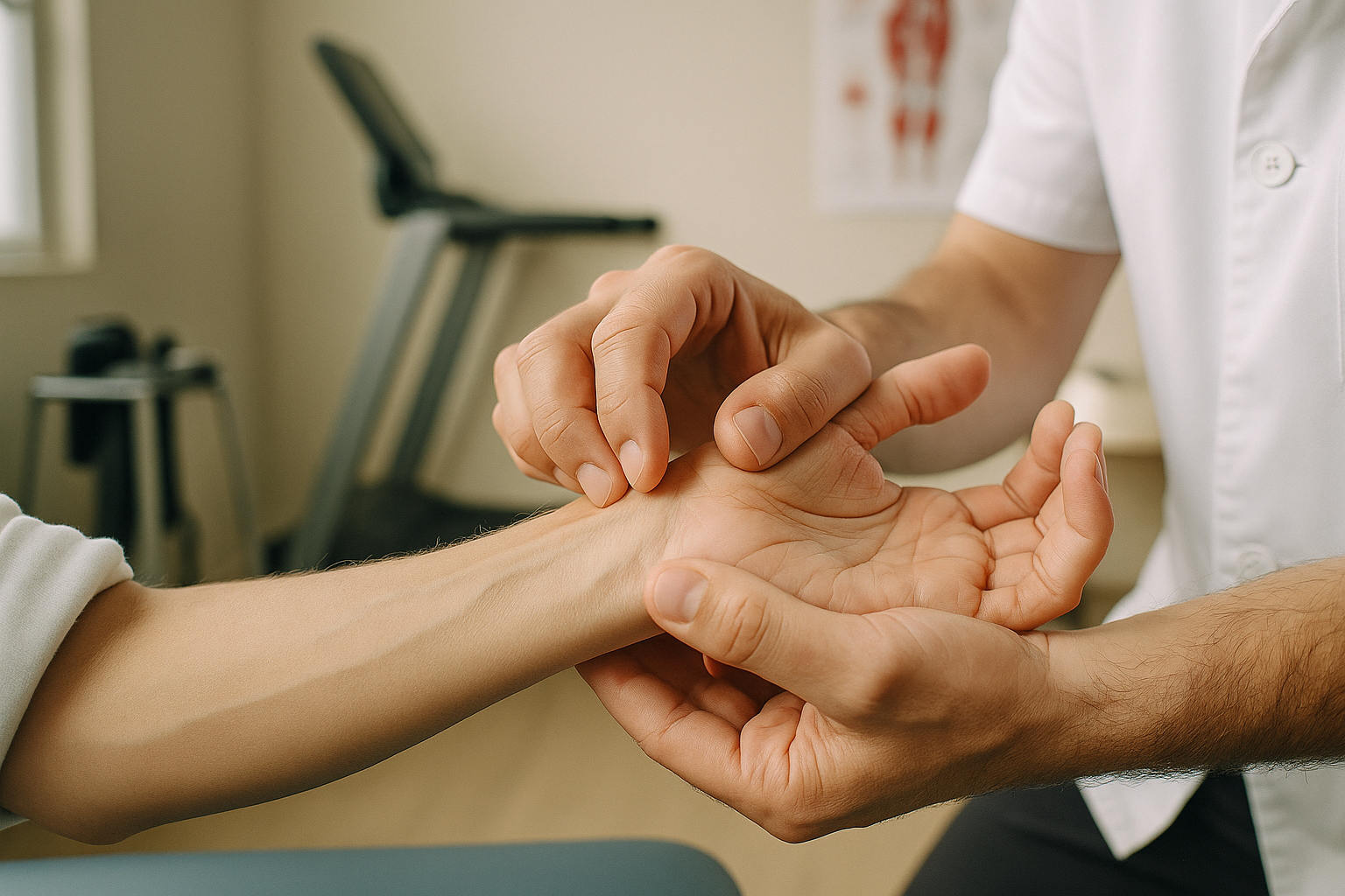
Physiotherapy Assessment: The Clinical Exam
The Importance of a Holistic Evaluation
A comprehensive evaluation is the foundation of successful recovery. Therefore, clinicians must adopt a holistic perspective immediately. We cannot simply focus on the wrist joint alone. Instead, we examine the entire upper kinetic chain. This includes checking the elbow, shoulder, and cervical spine.
Often, proximal issues manifest as distal symptoms in the hand. Consequently, ignoring these upstream areas leads to incomplete diagnoses. We also consider the patient’s daily activities. Do they perform repetitive tasks? Understanding their lifestyle helps pinpoint the root cause of injury. Thus, the assessment becomes a puzzle-solving process.
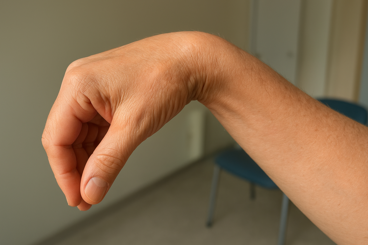
Visual Inspection and Posture
The physical exam starts the moment the patient enters the room. We carefully observe their natural resting position. For instance, is the wrist guarded tightly against the body? This protective posturing often signals acute trauma or severe inflammation. Additionally, we check for asymmetry between the hands.
Visible deformities or distinct swelling patterns provide immediate diagnostic data. We look for the following visual cues:
- Ganglion cysts: Often visible on the dorsal aspect.
- Muscle atrophy: Indicates nerve compression or disuse.
- Skin discoloration: Suggests vascular involvement or infection.
Furthermore, the position of the fingers can reveal tendon integrity. As a result, observation guides the subsequent hands-on examination.
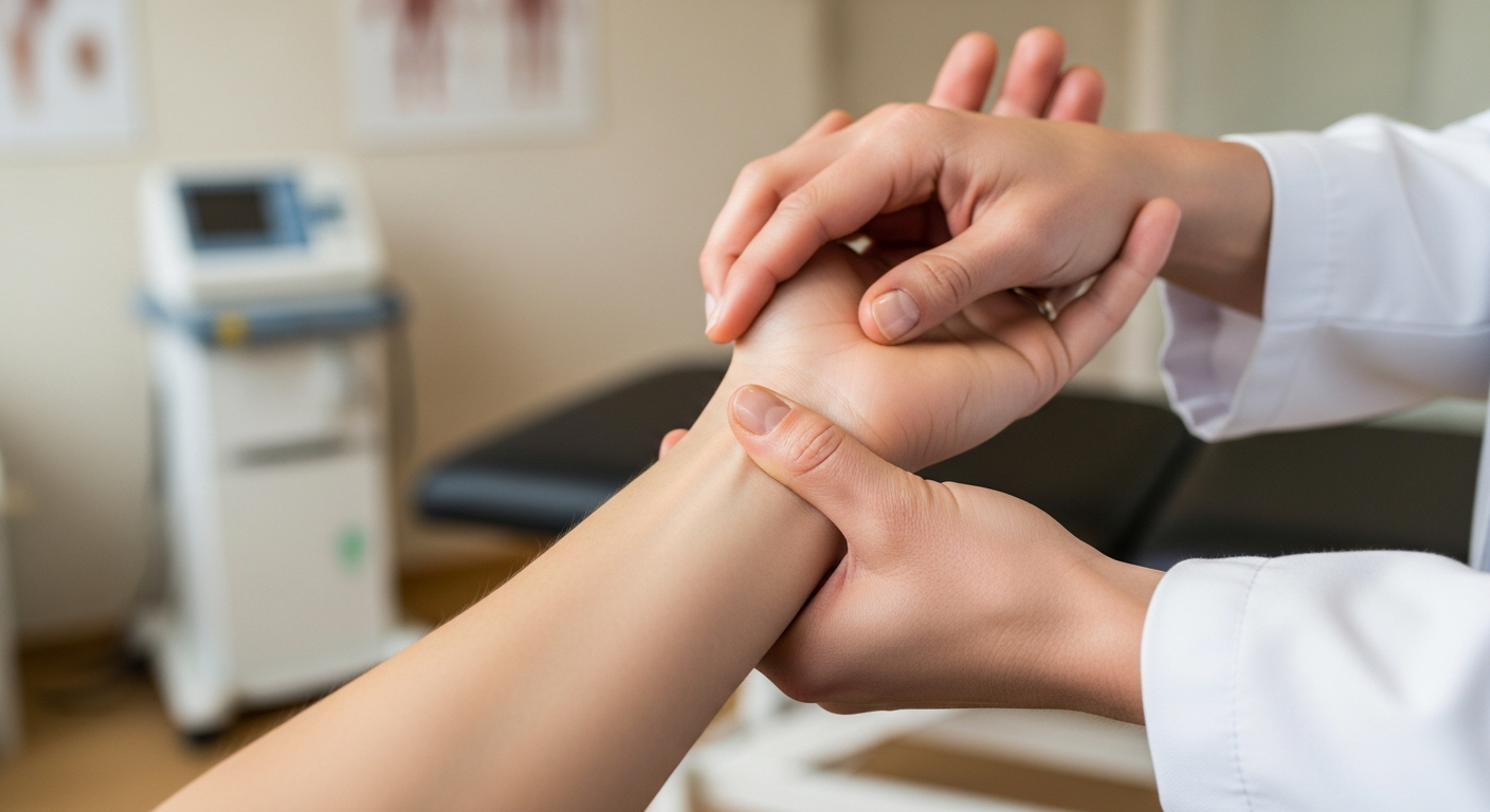
Precise Palpation Techniques
Hands-on assessment refines our differential diagnosis significantly. We systematically palpate the carpal bones to localize pain sources. Specifically, tenderness in the anatomical snuffbox is a critical red flag. As a result, this finding necessitates ruling out a scaphoid fracture immediately. Even minor discomfort here warrants imaging.
We also examine the pisiform and hook of the hamate. Meanwhile, assessing the soft tissue is equally vital. We trace the extensor tendons for signs of inflammation. This helps distinguish between bony pathology and conditions like De Quervain’s tenosynovitis. Therefore, precise touch maps the injury accurately.
Ulnar-Sided Pathologies
Pain on the pinky side of the wrist requires specific attention. Frequently, this indicates pathology within the Triangular Fibrocartilage Complex (TFCC). We test load-bearing capabilities in this specific region. Moreover, we assess joint stability during rotation.
A positive “fovea sign” often confirms deep ligamentous injury here. Consequently, distinct palpation separates tendonitis from structural tears. We also check the extensor carpi ulnaris (ECU) tendon. Is it snapping over the bone? Subluxation of this tendon is a common source of ulnar pain. Thus, differentiating these structures is essential for proper treatment.
Range of Motion and Strength
We measure active and passive range of motion next. Therapists use a goniometer for precision. We compare the injured side to the healthy side. Significant discrepancies indicate pathology. Additionally, we assess end-feel. A hard end-feel suggests bony blockage. A springy block might imply a meniscal or cartilage tear.
Strength testing follows mobility checks. We isolate specific muscle groups. Pain during resistance often points to tendinopathy. Conversely, weakness without pain might suggest a nerve injury. We must differentiate between the two carefully.
Special Tests for Instability
Clinical tests help us rule in specific conditions. For instance, the Watson scaphoid shift test detects scapholunate instability. The therapist applies pressure to the scaphoid while moving the wrist. A palpable “clunk” indicates a positive test. Similarly, the fovea sign tests for ulnar-sided issues. The examiner presses deep into the ulnar fovea. Sharp pain here usually implicates the TFCC.
However, these tests are not perfect. Sensitivity and specificity vary. Therefore, we correlate findings with patient history. Imaging often confirms our clinical suspicions. But the clinical exam remains the cornerstone of diagnosis.
Rehabilitation Strategies
Initial Assessment and Treatment Planning
When veterinarians pinpoint the specific carpal joint issue, the rehabilitation process starts immediately. Treatment objectives evolve based on where the patient falls within the injury timeline. For instance, a fresh injury requires different interventions than a weeks-old condition. Therefore, understanding the injury phase guides every clinical decision. Each stage demands unique approaches to optimize healing outcomes.
Acute Phase Management
During the acute phase, which typically spans the first 72 hours, protecting damaged structures takes priority. Source Consequently, practitioners implement strict protocols to prevent further harm. The joint experiences significant vulnerability during this critical window. Moreover, excessive movement can worsen tissue damage and prolong recovery time.
Immobilization Techniques
Veterinarians frequently apply custom-fitted splints to restrict carpal movement. These devices maintain the joint in optimal healing positions. For example, a palmar splint keeps the carpus in slight extension. Meanwhile, the immobilization prevents stress on injured ligaments or fractured bones. Additionally, splinting reduces pain by limiting motion at the injury site. However, the duration of immobilization varies based on injury severity. Most acute cases require one to three weeks of continuous support.
Inflammation Control Methods
Therapeutic modalities play a crucial role in managing acute inflammation. Ice therapy, or cryotherapy, remains the gold standard for initial treatment. Application of cold packs for 15-20 minute intervals reduces swelling effectively. Furthermore, cold therapy decreases metabolic activity in damaged tissues. As a result, secondary tissue damage diminishes significantly. Practitioners may also incorporate compression wraps alongside ice applications. This combination enhances fluid drainage from the affected area.
Restoring Mobility
Introducing Movement During Recovery
The transition from immobilization to active movement represents a critical phase in carpal joint rehabilitation. Gentle active range of motion exercises typically begin around week three to four, depending on tissue healing. These movements should feel comfortable and controlled. Patients learn to flex, extend, and rotate their wrist without forcing the motion. Furthermore, this gradual approach prevents re-injury while promoting healthy tissue adaptation.
Patient-Centered Movement Progression
We emphasize listening to the body’s signals during early mobilization. Pain serves as an important guide throughout this process. Consequently, patients should work within a pain-free range to avoid inflammatory responses. For example, if wrist extension causes discomfort at 30 degrees, we stop at 25 degrees. Additionally, we monitor for swelling or increased warmth after exercises. These signs indicate the need to modify activity levels.
Manual therapy Techniques
Manual therapy becomes increasingly valuable as tissues mature and strengthen. Joint mobilizations target specific restrictions within the carpal complex. Moreover, these hands-on techniques address stiffness that active movement alone cannot resolve. Therapists apply graded oscillations to the affected joints. The pressure varies from gentle to moderate based on patient tolerance.
Carpal Bone Gliding Techniques
The eight carpal bones require individual attention during manual treatment. Gliding mobilizations restore normal arthrokinematic motion between these small bones. For instance, the scaphoid may need posterior gliding to improve wrist extension. Meanwhile, the lunate might require anterior mobilization for better flexion mechanics. Therefore, each bone receives specific directional forces based on assessment findings.
Customized Treatment Approaches
Every carpal joint injury presents unique mechanical restrictions and healing patterns. Assessment guides our selection of appropriate manual techniques. However, we continuously reassess joint mobility throughout each session. As a result, treatment adapts to immediate tissue responses. Some patients need more aggressive mobilization, while others require gentler approaches. This individualized strategy optimizes outcomes and maintains patient comfort.
Strengthening and Proprioception
Finally, we focus on strength and stability. Isometric exercises offer a safe starting point. As tolerance improves, we add resistance. We use bands, weights, and functional tools. Furthermore, proprioception training is essential. The wrist relies on sensory feedback for stability. Exercises like holding a vibrating bar or rolling a ball on a wall challenge this system. Better proprioception prevents re-injury.
Conclusion
The wrist joint performs complex mechanical feats daily. Source Its intricate anatomy of bones, ligaments, and muscles allows for incredible versatility. However, this complexity creates vulnerability. Physiotherapists play a critical role in managing wrist dysfunction. Through careful assessment and targeted rehab, we restore function. Ultimately, a healthy wrist is essential for a functional life.
*As an Amazon Associate, I earn from qualifying purchases.