
Understanding Wrist X-Ray Imaging
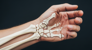
The wrist represents one of the most complex anatomical regions to evaluate through medical imaging. Source Radiologists and orthopedic specialists rely heavily on X-rays and CT scans to assess injuries and conditions affecting this intricate joint. Understanding how carpal bones appear on radiographic images helps clinicians identify fractures, dislocations, and alignment issues that might otherwise go undetected.
The Basics of Carpal Bone Anatomy
The wrist contains eight small carpal bones arranged in two rows, and each one has a distinct shape and position that radiographers and physicians must learn to recognize instantly. The proximal row (closest to the forearm) contains the scaphoid, lunate, triquetrum, and pisiform bones. The distal row (toward the hand) includes the trapezium, trapezoid, capitate, and hamate. Between these bones lie small joints and ligaments that allow for the wrist’s remarkable range of motion while maintaining stability.
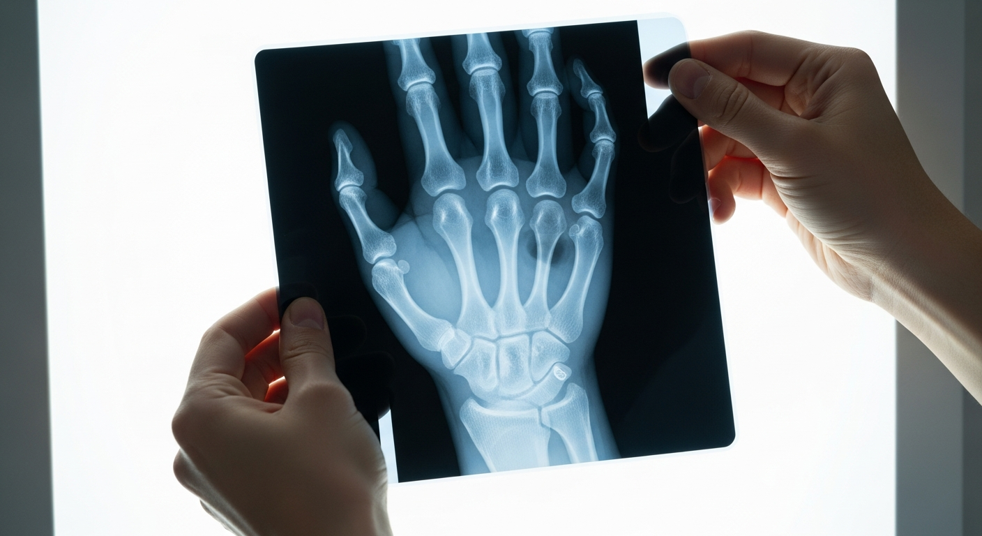
When viewing a standard posteroanterior (PA) wrist X-ray—taken with the palm facing down—each carpal bone casts a distinct shadow. The scaphoid appears as a boat-shaped bone on the thumb side, the lunate resembles a crescent moon in the center, and the triquetrum sits on the pinky side. Learning to identify these landmarks on your own radiographs takes practice, which is why having a physical reference tool can accelerate understanding considerably.
The Eight Carpal Bones: Names and Mnemonic
Learning the eight carpal bones is easier with a memory aid. Reading proximal-to-distal and radial-to-ulnar (from the thumb side inward), the classic mnemonic is “Some Lovers Try Positions That They Can’t Handle”:
- Scaphoid — the boat-shaped bone at the base of the thumb and the most commonly fractured carpal bone; its retrograde blood supply makes proximal-pole fractures prone to avascular necrosis if diagnosis is delayed.
- Lunate — the central, crescent-shaped bone that bears much of the axial load transmitted from hand to forearm and is the pivot point in perilunate dislocations.
- Triquetrum — a pyramid-shaped bone on the ulnar (pinky) side, frequently involved in dorsal chip fractures.
- Pisiform — a small sesamoid bone embedded in the flexor carpi ulnaris tendon; it overlaps the triquetrum and is best evaluated on the carpal tunnel or oblique view.
- Trapezium — forms the saddle joint at the base of the thumb; first carpometacarpal (CMC) arthritis here is among the most common forms of hand arthritis.
- Trapezoid — the smallest bone of the distal row, wedged tightly between its neighbors, so isolated fractures are uncommon.
- Capitate — the largest carpal bone and central keystone of the wrist, articulating with the concavity of the lunate at the midcarpal joint.
- Hamate — recognized by its palmar hook (the hamulus), it is vulnerable to hook fractures in athletes who grip bats, clubs, or racquets; CT is the gold standard when a hook fracture is suspected.
Standard X-Ray Views for the Carpal Bones
Going deeper: How to Identify Carpal Bones on X-Ray — a labeled, diagram-by-diagram walkthrough
A complete wrist series uses several projections, because no single angle shows all eight carpal bones clearly. Medical professionals typically order multiple views to build a complete picture, and understanding why your radiologist ordered each one helps you recognize that a thorough evaluation requires more than a single image.
PA (Posteroanterior) View
Taken with the palm facing down, the PA view is the standard starting point and the best projection for assessing bone alignment, the joint spaces between the carpal bones, and the distal radioulnar joint. On a well-positioned PA film the scaphoid, lunate, and triquetrum are seen clearly, and the smooth, unbroken arcs formed by the two carpal rows (Gilula’s arcs) can be traced for signs of dislocation.
Lateral View (the “Sagittal” Projection)
The lateral view photographs the wrist from the side, and it is what searchers often mean when they look for the “sagittal projection” of the carpal bones—it images the wrist in the sagittal plane. It is the essential view for assessing the alignment of the radius, lunate, and capitate: in a neutral wrist these three should stack in a straight longitudinal line, with the lunate seated squarely in the lunate fossa of the radius and the head of the capitate centered over the lunate. Loss of this colinear relationship is a key sign of carpal dislocation, such as a perilunate or lunate dislocation.
Oblique View
The oblique projection is taken like the PA view but with the wrist rotated roughly 45 degrees. It increases sensitivity to carpal, metacarpal, and phalangeal fractures and is particularly useful for the scaphoid tuberosity and for the trapezium and trapezoid, which overlap on other views.
Scaphoid (Ulnar-Deviation) View
The scaphoid is best seen on an elongated PA view taken with the wrist in about 10–15 degrees of ulnar deviation and the beam angled roughly 30 degrees toward the elbow. This position lengthens the scaphoid and opens up its waist, improving detection of the fractures that are so easily missed on a standard PA. Clenched-fist views can be added to show how the bones shift under a tight grip, which helps detect scapholunate ligament injuries.
Carpal Tunnel View
The carpal tunnel view is an axial projection taken with the wrist dorsiflexed so the carpals and metacarpals lift away from the film. It is especially helpful for injuries of the hook of the hamate, the pisiform, and the trapezium—bones that are frequently hidden by overlap on the routine views.
Identifying Common Wrist Injuries on X-Rays
Fractures of the distal radius (Colles’ fracture) are among the most frequent wrist injuries and typically show a distinctive break pattern on X-rays. Scaphoid fractures are trickier to spot initially because the fracture line may not be immediately visible on early radiographs, sometimes requiring follow-up imaging. Carpal dislocations—where bones shift out of their normal alignment—create obvious disruptions in the orderly arrangement of bone shadows.
Falls on an outstretched hand (the FOOSH mechanism) are the dominant cause of scaphoid fractures, and up to 20% are radiographically occult on the first film. Persistent tenderness in the anatomical snuffbox—the depression at the base of the thumb between the extensor pollicis longus and brevis tendons—warrants repeat imaging or MRI within about 10 to 14 days even when the initial X-ray looks normal, because a missed scaphoid fracture risks avascular necrosis. Perilunate dislocations are a true wrist emergency: on the lateral view the capitate is displaced dorsally while the lunate rotates palmarly, disrupting the normal radius–lunate–capitate line, and this pattern needs urgent surgical stabilization.
Arthritis of the wrist appears as progressive narrowing of the joint spaces between bones, along with bone spurs or osteophytes. Comparing old X-rays to new ones allows physicians to track whether degenerative changes are progressing. Ligament injuries themselves don’t show up on standard X-rays, but the bone misalignment they cause does, making radiographic analysis crucial for indirect assessment.
A 3D Model for Actually Understanding Wrist Anatomy Before Your Next Doctor Visit
If you’re dealing with wrist pain or a suspected carpal bone injury, holding a detailed anatomical model in your hands while looking at your own X-rays makes a massive difference in understanding what’s actually broken or misaligned. Most people sit through doctor appointments nodding along to terms like “scaphoid” and “lunate” without any real sense of where these bones are or how they move—this model changes that.
What works
- The removable bones let you see exactly how each carpal bone sits in relation to the radius and ulna, which makes X-ray images click immediately instead of looking like abstract shadows.
- Most people report that having a tactile reference cuts through medical jargon—you can actually point to the bone on the model and match it to your radiograph, giving you real context for your injury or condition.
- The hand-and-wrist assembly is at a scale that’s accurate enough to use alongside actual imaging reports, so you’re not learning from something that oversimplifies the anatomy.
What doesn’t
- This is a learning tool, not a diagnostic one—it won’t replace a radiologist’s reading, and if you’re using it to try to diagnose yourself instead of seeing a specialist, you’ll miss things.
- It’s static, so if your issue involves dynamic problems (like carpal instability during movement), a model alone won’t show you how your bones actually shift during gripping or rotation.
This works best as a companion to an actual medical consultation, not a replacement for one. EVOTECH SCIENTIFIC Hand anatomical model
When to Seek Professional Interpretation
While understanding basic wrist anatomy empowers you as a patient, radiographic interpretation requires specialized training that takes years to develop. Subtle fracture lines, early signs of arthritis, and ligament-related bone shifts can easily be missed by untrained eyes. If you suspect a wrist injury, an X-ray ordered by your physician and read by a radiologist remains the gold standard for diagnosis.
Bring questions to your appointments, ask your doctor to walk you through what they’re seeing on your images, and use learning tools to deepen your understanding of your own condition. This collaborative approach—combining professional expertise with your own anatomical literacy—leads to better treatment decisions and faster recovery.
Common Questions About Reading a Wrist X-Ray
Can you identify all eight carpal bones on a lateral X-ray?
Not reliably, and you are not supposed to. On a true lateral the carpal bones sit directly on top of one another, so they overlap into a dense cluster. What a lateral is actually for is alignment: a single straight axis should run through the middle of the radius, the lunate and the capitate. The lunate should look like a cup sitting on the radius with the round head of the capitate resting inside it — the “cup within a cup” appearance. The scaphoid and the pisiform are the two that project forward, toward the palm. If you want to name individual bones, the PA view is the one to use.
What is normal spacing between the carpal bones?
Roughly one to two millimetres, and — just as importantly — even all the way across. The scapholunate gap specifically should be 2 mm or less. Radiologists also check that Gilula’s three arcs run smooth and unbroken across the carpus; a step or break in an arc means a bone has shifted out of position, even when nothing looks obviously fractured.
How can I tell which bone is which on my own X-ray?
Find the capitate first. It is the largest carpal bone and it sits dead centre, at the base of the middle finger, so everything else can be placed relative to it. Then work along each row from the thumb side: proximal row is scaphoid, lunate, triquetrum, pisiform; distal row is trapezium, trapezoid, capitate, hamate. The old mnemonic — Some Lovers Try Positions That They Cannot Handle — runs in exactly that order.
This is general information to help you follow along with your own care, not medical advice — your own clinician has examined you and this page has not.
About This Article

Written by Michael Torres. Michael Torres has had joint problems his whole life — hypermobile as a kid, chronic sprains and a dislocating shoulder through his twenties, an osteoarthritis diagnosis in his early fifties, and a right knee replacement since. He is not a doctor or a physical therapist and has no clinical training. He writes about what has helped him and what he has watched help the people around him, and says plainly when he has not tried something himself. More about this site.
Scope and limits. Michael has no medical or clinical training — he writes about living with his own joint pain, not about treating anyone else's. Nothing here is medical advice. Talk to a doctor before starting anything new, and get seen promptly for pain that is new, severe, worsening, or comes with swelling, fever, or numbness.
Verify this against a primary source
Specifics change, and your situation may differ from the one described here. These are the authorities worth checking directly:
- NIAMS — arthritis and musculoskeletal research from the NIH
- MedlinePlus — plain-language medical reference from the NIH
- Arthritis Foundation — condition guides and treatment overviews