Understanding Anatomy and Function
Understanding the Complex Architecture of the Wrist
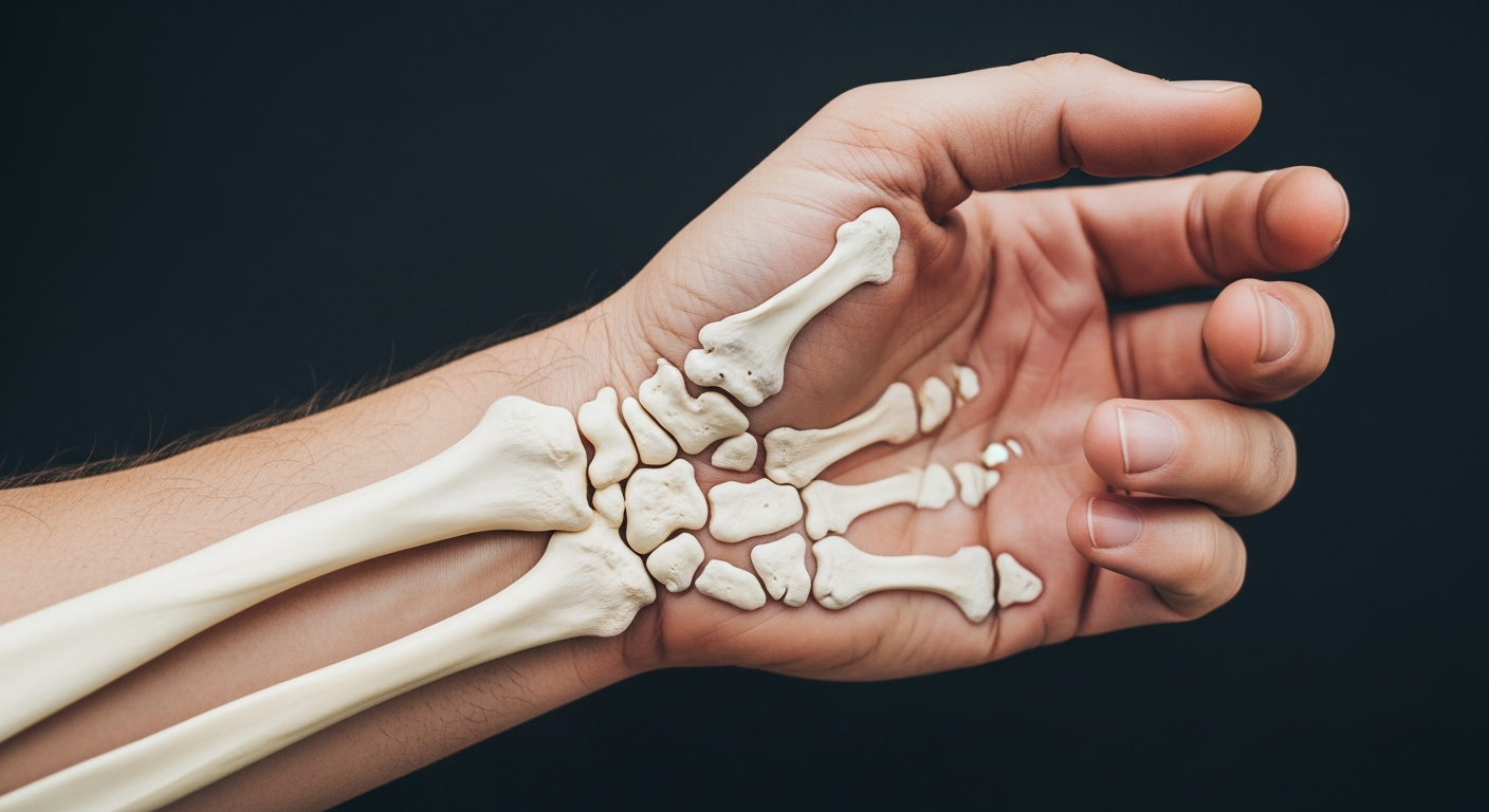
The wrist represents one of the most sophisticated joint systems in the human body. Furthermore, this remarkable structure facilitates everything from delicate finger movements to powerful gripping actions. Most people never consider the complexity hidden beneath their skin. However, the wrist contains an elaborate network of bones, ligaments, and tendons working in perfect harmony.
When dealing with wrist and hand discomfort from repetitive activities like typing or extended travel documentation work, having the right supportive gear can make all the difference in maintaining comfort throughout your adventures. A quality wrist brace provides essential stabilization and copper-infused support that can help reduce inflammation during long writing sessions about your travel experiences. For travelers who spend hours typing blog posts or editing photos, investing in compression gloves offers fingerless freedom while still providing crucial support to reduce strain from repetitive keyboard use. Creating a comfortable workspace while traveling becomes much easier when you add a wrist rest that cushions your arms with memory foam during those marathon editing sessions in hotel rooms or cafes. Many digital nomads and travel bloggers find that compression gloves designed for all-around support help them maintain productivity without sacrificing hand comfort during their journeys around the world. For comprehensive hand care solutions while traveling, exploring options from medical supply specialists can provide professional-grade products that address various wrist and hand concerns you might encounter on the road. When inflammation or acute pain strikes after a long day of photography or hiking, a therapeutic ice pack wrap designed specifically for wrists can provide soothing hot and cold therapy right in your accommodation. Pairing your keyboard setup with an ergonomic mouse pad featuring gel wrist support helps prevent strain during those long hours of travel planning and content creation. Building hand and forearm strength with a grip strengthener kit can actually help prevent injuries before they occur, especially important for travelers who carry heavy camera equipment or backpacks regularly. A dedicated wrist splint designed for nighttime wear ensures your wrists heal properly while you sleep, so you wake up refreshed and ready for another day of adventure. Finally, keeping a pair of compression gloves specifically designed by medical professionals in your travel first aid kit ensures you’re always prepared to address hand discomfort wherever your journeys take you around the globe.
The carpal bones serve as the foundation for all wrist movement. These eight small bones are roughly the size of large peas or small marbles. Moreover, they arrange themselves in two parallel rows within the wrist joint. This unique configuration creates a flexible yet stable platform. As a result, your hand can move in multiple directions simultaneously.
The proximal row sits closest to your forearm bones. Meanwhile, the distal row connects directly to your hand bones. Additionally, this dual-row arrangement functions like a universal joint. Therefore, you can bend, extend, and rotate your wrist with remarkable precision. Each bone plays a specific role in this mechanical masterpiece.
The Importance of Carpal Joint Health
Daily activities place tremendous demands on these tiny structures. For instance, typing on a keyboard requires thousands of small movements. Similarly, lifting a coffee cup involves precise coordination between all eight bones. Consequently, understanding carpal anatomy becomes essential for maintaining hand health. Furthermore, this knowledge helps identify potential problems before they become serious.
Many conditions can affect the carpal region throughout life. Carpal tunnel syndrome ranks among the most common complaints. However, fractures, arthritis, and ligament injuries also frequently occur. Therefore, learning about these structures empowers better health decisions. Moreover, early recognition of symptoms can prevent long-term damage.
The Proximal Row of Carpal Bones
The proximal row of carpal bones consists of four bones situated closest to the forearm. These bones include the scaphoid, lunate, triquetrum, and pisiform. Each bone plays a crucial role in wrist movement and stability.
Scaphoid Bone
The Scaphoid’s Anatomical Significance
The scaphoid bone stands out as the most substantial bone within the proximal carpal row. Moreover, it occupies a critical position in the wrist’s structural framework. This boat-shaped bone derives its name from the Greek word “skaphe,” meaning boat. Additionally, its unique curved shape allows it to bridge two important carpal rows.
The scaphoid’s location places it on the radial side of the wrist. Consequently, it sits directly beneath the thumb’s base. Furthermore, this positioning makes it particularly vulnerable during falls. The bone forms a crucial articulation with the radius bone. Therefore, it serves as a primary connection point between the forearm and hand.
Functional Role in Wrist Mechanics
The scaphoid functions as a mechanical stabilizer during wrist movements. For example, it prevents excessive motion during gripping activities. Additionally, it transfers forces from the hand to the forearm. The bone acts like a keystone in an arch. As a result, it maintains proper alignment of surrounding carpal bones.
During wrist flexion and extension, the scaphoid rotates significantly. Meanwhile, it also shifts position during radial and ulnar deviation. This dynamic movement pattern makes it essential for complex hand motions. Furthermore, activities like throwing, pushing, or lifting rely heavily on scaphoid stability. Without proper scaphoid function, the wrist loses considerable strength and mobility.
Lunate Bone
The Lunate’s Strategic Position
The lunate bone occupies a critical central location within the carpal joint architecture. Moreover, its crescent-shaped structure sits directly adjacent to the scaphoid bone. This positioning makes it a cornerstone of wrist stability. The lunate forms essential connections with multiple surrounding bones. Specifically, it articulates with the radius bone above it. Additionally, it connects to the capitate bone below. Source Therefore, the lunate serves as a vital link in the wrist’s complex mechanical system.
Primary Functions in Wrist Movement
The lunate operates as the primary pivot point for hand movements. Consequently, every time you bend your wrist forward or backward, the lunate coordinates this action. Furthermore, the bone facilitates smooth flexion when you curl your hand downward. Similarly, it enables extension when you tilt your hand upward. For example, typing on a keyboard requires constant lunate articulation. The bone’s curved shape allows it to rock back and forth efficiently. As a result, the lunate distributes forces evenly across the wrist joint.
Clinical Significance
Kienböck’s disease specifically affects the lunate bone’s blood supply. However, this condition can lead to bone deterioration over time. Additionally, lunate fractures often occur from falls onto an outstretched hand. Therefore, medical professionals closely monitor lunate health during wrist examinations. The bone’s central position makes it vulnerable to repetitive stress injuries.
Triquetrum Bone
Position and Location of the Triquetrum
The triquetrum bone occupies a strategic position within the carpal architecture. It sits along the ulnar border of the wrist. This placement makes it the most medial bone in the proximal carpal row. Furthermore, Source the triquetrum forms a crucial bridge between multiple carpal structures. It articulates directly with the lunate bone on its radial side. Additionally, it connects with the pisiform bone anteriorly. The hamate bone sits just below it in the distal carpal row.
Structural Support Function
The triquetrum serves as a primary stabilizer for the ulnar wrist region. This small pyramidal bone distributes forces from the hand to the forearm. Moreover, it acts as a keystone in maintaining proper carpal alignment. The bone’s unique shape allows it to interlock with surrounding structures. Consequently, this interlocking mechanism prevents excessive lateral movement. The triquetrum also helps absorb impact during weight-bearing activities.
Clinical Significance
Healthcare professionals recognize the triquetrum’s importance in wrist mechanics. For example, it frequently experiences stress fractures in athletes. Additionally, the bone can dislocate during traumatic injuries. Therefore, proper assessment of this carpal bone is essential. Imaging studies often focus on triquetrum positioning during diagnostic evaluations. Meanwhile, surgeons must consider its role when planning wrist reconstruction procedures.
Pisiform Bone
Unique Anatomical Classification
The pisiform stands out anatomically because it is technically a sesamoid bone. This means it develops entirely within a tendon. Specifically, it sits inside the flexor carpi ulnaris tendon. Consequently, it functions differently than its neighbors in the proximal row. Other carpal bones articulate directly to transfer axial loads. In contrast, the pisiform acts primarily as a pulley system. This structure provides a smooth, gliding surface for tendon movement. Therefore, it is distinct from the standard load-bearing carpal architecture. Source ## Structural Relationships and Location
You can easily locate this bone on the palmar side of the wrist. It rests at the base of the small finger. Furthermore, it articulates solely with the triquetrum bone. This unique connection creates the pisotriquetral joint. As a result, this plane joint allows for subtle gliding movements. These motions are crucial for overall wrist flexibility during complex tasks.
Key anatomical neighbors include:
- Triquetrum: The bone directly beneath the pisiform.
- Hamate: Located distally to the pisiform.
- Flexor Carpi Ulnaris: The muscle tendon encasing the bone.
Biomechanical Functions
Its primary function involves mechanical leverage. By projecting outward, the bone acts as a spacer. Therefore, it holds the flexor tendon away from the wrist’s center of rotation. This increases the force generated during wrist flexion. Without this bone, grip strength would likely decrease.
Additionally, the pisiform serves a vital structural role. It forms the medial border of Guyon’s canal. Thus, it helps protect the passing ulnar nerve and artery. Moreover, it anchors the transverse carpal ligament. This ligament forms the roof of the carpal tunnel. Consequently, the pisiform is essential for stabilizing the hand’s soft tissues.
The Distal Row of Carpal Bones
Understanding the distal carpal row Structure
The distal carpal row forms the foundation for metacarpal bone attachment. This row creates a stable platform for finger movement. Furthermore, these four bones work together as an integrated unit. Each bone has distinct characteristics that contribute to overall hand mechanics.
The trapezium sits at the thumb side of this row. It features a unique saddle-shaped joint surface. This configuration allows the thumb its remarkable range of motion. Additionally, the trapezium articulates with the first metacarpal bone. This connection enables opposition movements essential for gripping objects.
Individual Bone Functions
The trapezoid lies adjacent to the trapezium in the sequence. This smaller bone connects with the second metacarpal. Moreover, it provides stability to the index finger’s base. The trapezoid acts as a critical anchor point during precision tasks.
The capitate represents the largest carpal bone in the entire wrist. It occupies the central position within the distal row. Consequently, this bone bears significant load during hand activities. The capitate articulates with the third metacarpal bone. Therefore, it directly influences middle finger function and strength.
The Hamate’s Distinctive Features
The hamate completes the distal row on the pinky finger side. This bone possesses a distinctive hook-like projection called the hamulus. Additionally, the hamate connects with both fourth and fifth metacarpals. This dual connection provides support for the ring and pinky fingers.
These bones collectively form the carpometacarpal joints. These articulations allow controlled finger movements during daily activities. For example, writing, typing, and tool manipulation all depend on these connections. Furthermore, the distal row transfers forces from the fingers to the forearm. This force transmission occurs during pushing, pulling, and lifting movements.
Clinical Significance in Hand Function
The positioning of these bones near the fingers creates functional advantages. They enable fine motor control necessary for delicate tasks. Meanwhile, they maintain structural integrity during power grips. This dual capability makes the distal row essential for hand versatility.
Injuries to these bones can significantly impair hand function. Fractures often result from falls onto an outstretched hand. However, proper diagnosis and treatment typically restore normal function. Understanding their anatomy helps clinicians develop effective rehabilitation protocols.
Trapezium Bone
Located near the base of the thumb, the trapezium allows for thumb rotation and opposition. This bone is essential for grasping and pinching motions.
Trapezoid Bone

Anatomical Structure and Location
The trapezoid bone is the smallest element within the distal carpal row. It features a distinctive wedge shape with a broader dorsal surface. Specifically, this unique orientation helps maintain the transverse carpal arch. Consequently, it provides essential structural stability to the wrist complex.
Furthermore, this bone is securely locked into position by strong ligaments. It articulates with four neighboring bones to distribute physical stress effectively. These connections prevent excessive movement during heavy lifting or impact. Therefore, the wrist remains aligned even under significant pressure.
Its specific articulations include:
- Proximally: Connects with the scaphoid.
- Distally: Anchors the second metacarpal.
- Laterally: Borders the trapezium and capitate.
Functional Importance
The primary role of the trapezoid is anchoring the second metacarpal. This is the long bone connecting directly to the index finger. As a result, it creates a relatively immobile joint compared to the thumb. This rigidity is vital for tasks requiring steadiness and precision.
For example, the trapezoid facilitates several key actions:
- Pinching: It provides a firm base for the index finger to press against the thumb.
- Key Grips: It stabilizes the hand when turning a key or holding a credit card.
- Impact Absorption: It transfers force from the index finger through the wrist.
Additionally, the trapezoid transmits force from the hand to the forearm. Without this transmission, the wrist would suffer from significant instability. Thus, even minor injuries to this area can disrupt overall hand function.
Clinical Significance
Due to its protected position, fractures of the trapezoid are relatively rare. The bone is surrounded by a sturdy shell of other carpals. However, high-impact trauma can still cause damage to this area. For instance, a direct blow to the second metacarpal can transmit force backward.
Diagnosing these injuries often requires advanced imaging techniques. Standard X-rays may not show the fracture clearly due to bone overlap. Therefore, doctors frequently utilize CT scans for accurate assessment. Moreover, untreated injuries can lead to long-term arthritis in the joint.
Capitate Bone
The Capitate’s Dominant Position
The capitate bone stands out as the wrist’s most substantial carpal element. Moreover, Source it occupies a strategic central location within the carpal architecture. This positioning places it at the heart of wrist mechanics. The bone measures approximately 20-25 millimeters in length. Furthermore, its rounded head gives the capitate its distinctive name. The term “capitate” derives from the Latin word for “head.”
Structural Role in Wrist Stability
The capitate functions as the wrist’s primary stabilizing anchor. Additionally, it bears significant compressive forces during hand movements. The bone connects directly with the third metacarpal. This connection creates a rigid central column for the hand. Therefore, the capitate distributes loads efficiently across the wrist. It acts like a cornerstone in architectural design. Without this central support, wrist integrity would be compromised.
Keystone Function in Articulation
As a mechanical keystone, the capitate coordinates multiple joint surfaces. It articulates with seven different bones in total. For example, it contacts the scaphoid and lunate proximally. Distally, it connects with the second, third, and fourth metacarpals. Consequently, this extensive network enables complex wrist movements. The capitate facilitates flexion, extension, and radial deviation. Meanwhile, it maintains structural cohesion during these motions. This dual role makes it irreplaceable in wrist biomechanics.
Hamate Bone
Anatomical Position and Structure
The hamate bone occupies a distinctive position within the wrist’s architecture. Specifically, it sits on the ulnar side, which corresponds to the pinky finger side of your hand. This strategic placement makes it the fourth bone in the distal carpal row. Furthermore, its location allows it to interact with multiple surrounding bones, creating crucial joint surfaces.
The most recognizable feature is the hook of hamate, a curved, bony projection. This hook extends from the palmar surface of the bone. Moreover, it can be felt through the skin at the base of your palm. The projection measures approximately 1-2 centimeters in length. Source Additionally, its shape resembles a fishing hook or a bent finger pointing toward the thumb.
Muscular and Ligamentous Attachments
The hamate serves as a vital attachment site for numerous soft tissue structures. For example, the flexor digiti minimi brevis muscle originates from this bone. This muscle controls movement of your little finger. Meanwhile, the opponens digiti minimi also attaches here, enabling opposition movements.
Several important ligaments anchor to the hamate’s surfaces. The pisohamate ligament connects it to the pisiform bone nearby. Additionally, the transverse carpal ligament attaches to the hook of hamate. This ligament forms the roof of the carpal tunnel. Consequently, it protects the median nerve and flexor tendons passing beneath.
Functional Contributions to Grip
The hamate’s anatomical features directly enhance hand function and grip capabilities. Its hook provides mechanical advantage for the small finger flexor muscles. Therefore, activities requiring power grip benefit from this structure. For instance, holding a baseball bat or golf club relies on these attachments.
Furthermore, the bone acts as a pulley for flexor tendons. These tendons glide around the hook during finger movements. As a result, the hamate contributes to efficient force transmission. Moreover, it stabilizes the ulnar side of the palm during gripping activities.
Function of the Wrist as a Gliding Joint
The Anatomy of Gliding
The human wrist is a marvel of biological engineering. Specifically, it consists of eight small carpal bones. These bones are arranged in two distinct rows. Furthermore, they interact to create a unique gliding mechanism. Unlike simple hinges, these flat surfaces slide past one another smoothly. Consequently, this design reduces friction during repetitive actions. The bones are held together by strong ligaments. These ligaments provide essential stability. However, they still permit a fluid range of motion.
Mechanics of Movement
This anatomical arrangement enables four primary directional movements. Source Each movement serves a specific function in daily life. Additionally, these actions often combine for complex tasks.
The core movements include:
- Flexion: This occurs when you bend your palm downward.
- Extension: Conversely, this happens when you pull your hand back.
- Radial Deviation: This moves the hand toward the thumb side.
- Ulnar Deviation: Meanwhile, this shifts the hand toward the little finger.
Therefore, the wrist is highly adaptable. It can position the hand in almost any orientation.
Functional Importance
These movements are vital for executing fine motor skills. Without them, dexterity would be nearly impossible. For example, typing on a keyboard requires rapid, micro-adjustments. Similarly, gripping a pen demands both stability and flexibility. Thus, the carpal joint bridges the gap between power and precision. It transforms raw arm strength into delicate finger actions. Moreover, this versatility extends to artistic endeavors.
Consider the complexity of playing a musical instrument. A pianist’s wrists must glide effortlessly across the keys. Furthermore, athletes use this joint for ball control. A basketball player snaps their wrist to shoot. As a result, the carpal bones often absorb significant stress. Ultimately, this joint is essential for human interaction with the world.
Common Wrist Issues: Carpal Tunnel Syndrome and Fractures
The wrist’s complexity makes it susceptible to various conditions. Source Carpal Tunnel Syndrome (CTS) is one of the most common issues affecting the wrist. It occurs when the median nerve becomes compressed, leading to pain, tingling, and numbness in the hand. CTS often results from repetitive motions, such as typing or assembly work .
Understanding Scaphoid fractures
Scaphoid fractures represent one of the most common wrist injuries among active individuals. The scaphoid bone sits on the thumb side of the wrist. It connects the two rows of carpal bones together. When someone falls forward, they instinctively extend their hand to break the fall. This natural protective reflex places tremendous force directly on the scaphoid. Consequently, the bone can crack or break completely under this sudden pressure.
High-Risk Groups and Activities
Certain populations face elevated risks for these specific injuries. For example, athletes participating in contact sports experience frequent wrist trauma. Skateboarders, snowboarders, and cyclists commonly sustain scaphoid injuries during falls. Additionally, older adults with reduced bone density are particularly vulnerable. Young men between ages 20 and 30 represent the highest incidence group. Meanwhile, workplace accidents involving falls also contribute significantly to scaphoid fracture statistics.
Why Immediate Treatment Matters
The scaphoid bone has limited blood supply compared to other wrist bones. Therefore, delayed diagnosis can lead to serious long-term problems. Avascular necrosis may develop when blood flow becomes compromised. This condition causes bone tissue death and permanent damage. Furthermore, untreated fractures often result in nonunion, meaning the bone fails to heal. Patients may then develop chronic pain and arthritis. However, early intervention dramatically improves healing outcomes and prevents these complications.
Recognizing the Warning Signs
Several symptoms indicate a possible scaphoid fracture requiring evaluation:
- Tenderness in the anatomical snuffbox area near the thumb base
- Swelling along the thumb side of the wrist
- Pain that worsens when gripping or pinching objects
- Decreased range of motion in the wrist joint
- Bruising that appears hours after the initial injury
Moreover, some fractures produce subtle symptoms that patients initially dismiss. As a result, many individuals delay seeking medical care for several days or weeks.
Diagnostic Procedures
Physicians use multiple imaging techniques to confirm scaphoid fractures. Standard X-rays serve as the first diagnostic tool. However, these images may not reveal hairline fractures immediately after injury. Consequently, doctors often order MRI scans or CT scans for definitive diagnosis. In some cases, repeat X-rays taken 10-14 days later show fractures more clearly. Meanwhile, bone scans can detect stress fractures that other methods miss.
Treatment Approaches and Recovery
Treatment protocols vary based on fracture location and severity. Non-displaced fractures typically require cast immobilization for 8-12 weeks. The cast often extends to include the thumb for proper stabilization. Conversely, displaced fractures usually necessitate surgical intervention. Surgeons insert small screws to hold bone fragments in correct alignment. Additionally, physical therapy becomes essential after cast removal. Patients gradually rebuild strength and restore full wrist mobility. Therefore, complete recovery may take anywhere from three to six months depending on individual factors.
Identifying Carpal Bones: Simplified Diagrams
For those interested in anatomy, simplified diagrams of the wrist can aid in identifying the carpal bones. These visual aids often highlight each bone’s location and function, making it easier to understand their roles in wrist mechanics.
Conclusion
In conclusion, the carpal bones are integral to the wrist’s functionality and versatility. Understanding their anatomy and function helps us appreciate the complexity behind everyday hand movements. By recognizing common wrist issues, individuals can take preventive measures to maintain wrist health. Whether you’re an anatomy enthusiast or simply curious, exploring the intricacies of the carpal bones can deepen your appreciation for the human body’s design.
When dealing with wrist and hand discomfort from repetitive activities like typing or extended travel documentation work, having the right supportive gear can make all the difference in maintaining comfort throughout your adventures. A quality wrist brace provides essential stabilization and copper-infused support that can help reduce inflammation during long writing sessions about your travel experiences. For travelers who spend hours typing blog posts or editing photos, investing in compression gloves offers fingerless freedom while still providing crucial support to reduce strain from repetitive keyboard use. Creating a comfortable workspace while traveling becomes much easier when you add a wrist rest that cushions your arms with memory foam during those marathon editing sessions in hotel rooms or cafes. Many digital nomads and travel bloggers find that compression gloves designed for all-around support help them maintain productivity without sacrificing hand comfort during their journeys around the world. For comprehensive hand care solutions while traveling, exploring options from medical supply specialists can provide professional-grade products that address various wrist and hand concerns you might encounter on the road. When inflammation or acute pain strikes after a long day of photography or hiking, a therapeutic ice pack wrap designed specifically for wrists can provide soothing hot and cold therapy right in your accommodation. Pairing your keyboard setup with an ergonomic mouse pad featuring gel wrist support helps prevent strain during those long hours of travel planning and content creation. Building hand and forearm strength with a grip strengthener kit can actually help prevent injuries before they occur, especially important for travelers who carry heavy camera equipment or backpacks regularly. A dedicated wrist splint designed for nighttime wear ensures your wrists heal properly while you sleep, so you wake up refreshed and ready for another day of adventure. Finally, keeping a pair of compression gloves specifically designed by medical professionals in your travel first aid kit ensures you’re always prepared to address hand discomfort wherever your journeys take you around the globe.
*As an Amazon Associate, I earn from qualifying purchases.