Quick Summary: Carpal Bone Anatomy on Wrist X-Ray
- Eight carpal bones are arranged in two rows between the forearm and the metacarpals, forming one of the most mechanically complex joints in the human body.
- The proximal row — scaphoid, lunate, triquetrum, and pisiform — transmits force directly from the hand to the radius and ulna.
- According to the American Academy of Orthopaedic Surgeons (AAOS), scaphoid fractures are the most commonly missed carpal injuries on initial X-ray due to their occult presentation in up to 20% of cases.
- Perilunate dislocations represent a wrist emergency; the disrupted cup-and-ball alignment of the radius, lunate, and capitate is identifiable on a lateral X-ray view.
- Plain X-ray remains the essential first-line imaging tool after wrist trauma, but MRI or CT is often required to confirm ligament injuries and occult fractures.
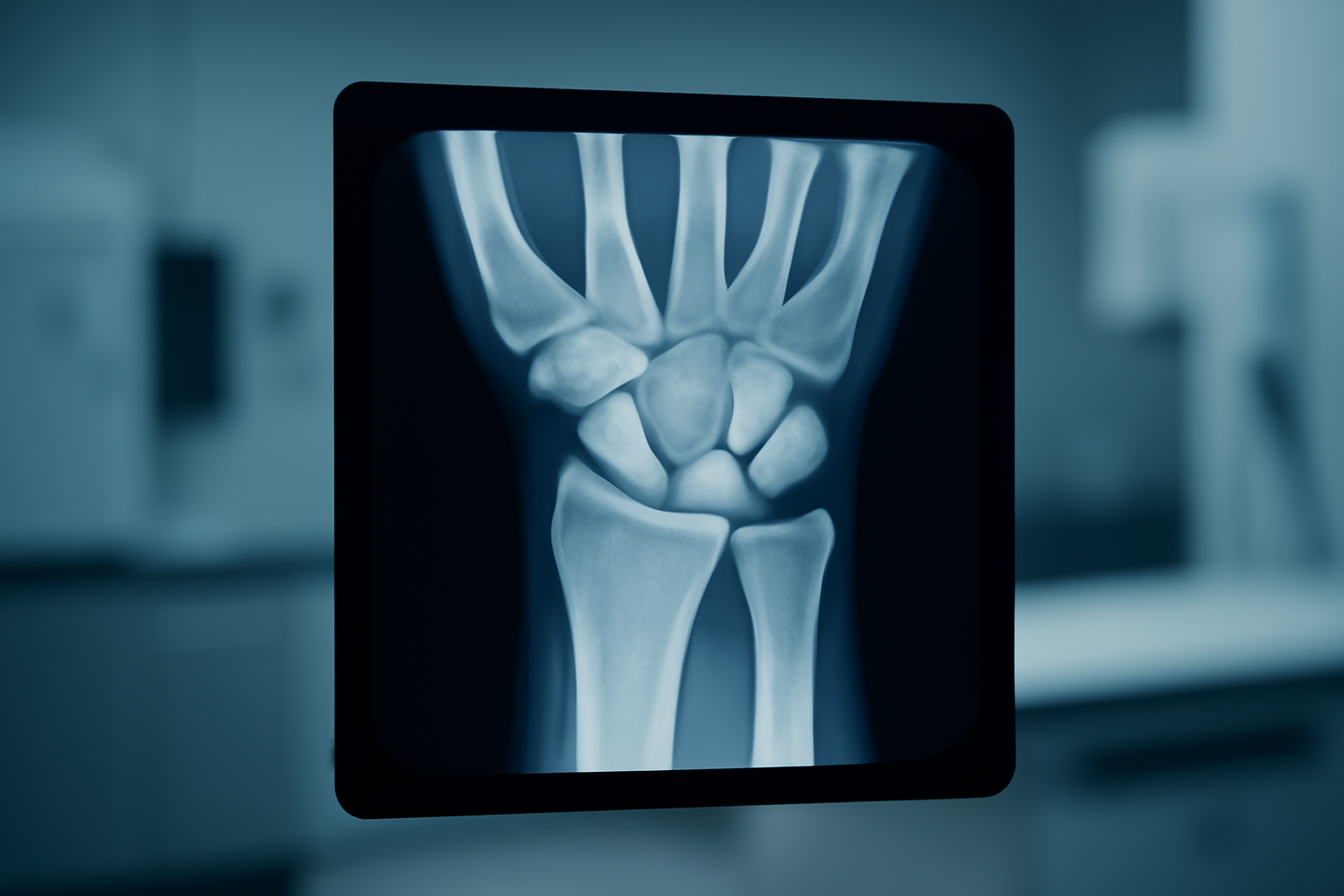
What Are the Eight Carpal Bones Visible on a Wrist X-Ray?
The wrist is one of the most architecturally complex joints in the human body. Sandwiched between the forearm bones — the radius and ulna — and the five metacarpals sit eight small carpal bones arranged in two functional rows. When patients review their wrist X-ray, orienting around these two rows is the most reliable starting point. JointHealthFAQ’s clinical review team, working with board-certified physical therapists and orthopedic specialists, has structured the following breakdown to help patients and caregivers understand exactly what they are looking at.
The Proximal Row: Bones Closest to the Forearm
Reading from the thumb side inward, the proximal row contains four bones:
- Scaphoid — The most commonly fractured carpal bone, the scaphoid sits at the base of the thumb and displays a distinctive curved, boat-like shape on X-ray. Its retrograde blood supply — entering distally and traveling proximally — makes proximal pole fractures particularly vulnerable to avascular necrosis if diagnosis is delayed.
- Lunate — Centrally positioned and crescent-shaped, the lunate bears a substantial proportion of axial load transmitted from hand to forearm. Research published in the Journal of Hand Surgery identifies lunate dislocations as a high-energy injury pattern in which the normal colinear alignment of the radius, lunate, and capitate is visibly disrupted on the lateral X-ray view.
- Triquetrum — Located on the ulnar side of the proximal row, this pyramid-shaped bone is frequently involved in dorsal chip fractures and interosseous ligament injuries that may appear subtle on standard posteroanterior (PA) views.
- Pisiform — A sesamoid bone embedded within the flexor carpi ulnaris tendon, the pisiform overlaps the triquetrum on certain projections and is best evaluated on the carpal tunnel or oblique view.
The Distal Row: Bones Closest to the Hand
The distal row is more rigidly interlocked and contributes to the stable foundation of the hand:
- Trapezium — Forming the saddle joint at the base of the thumb, the trapezium enables the thumb’s unique circumduction. The AAOS notes that first carpometacarpal (CMC) arthritis — visible as joint space narrowing at the trapezium — is among the most prevalent forms of hand arthritis, particularly in individuals over 50.
- Trapezoid — The smallest bone in the distal row, the trapezoid is wedge-shaped and tightly constrained by its neighbors, making isolated fractures uncommon.
- Capitate — The largest carpal bone and the central keystone of the wrist, the capitate has a rounded head that articulates with the concavity of the lunate, forming the critical midcarpal joint.
- Hamate — Recognized by its palmar hook projection (the hamulus), the hamate is susceptible to hook fractures in athletes who grip clubs, bats, or racquets. Standard PA views frequently miss this injury; CT imaging is the diagnostic gold standard when clinical suspicion is high.
A widely used mnemonic for the eight carpal bones in proximal-to-distal, radial-to-ulnar order: “Some Lovers Try Positions That They Can’t Handle” — Scaphoid, Lunate, Triquetrum, Pisiform, Trapezium, Trapezoid, Capitate, Hamate.
Which Wrist Injuries Are Most Visible on X-Ray?
Plain radiography remains the essential first-line imaging modality after wrist trauma. Clinicians working in hand surgery and emergency settings consistently evaluate the following injury patterns on initial X-ray series.
Scaphoid Fractures and the FOOSH Mechanism
Falls on an outstretched hand (FOOSH) are the dominant mechanism for scaphoid fractures. According to the AAOS, up to 20% of scaphoid fractures appear radiographically occult on initial imaging. Orthopedic specialists and licensed occupational therapists in clinical practice consistently note that persistent tenderness in the anatomical snuffbox — the depression between the extensor pollicis longus and extensor pollicis brevis tendons at the thumb base — warrants repeat imaging or MRI within 10 to 14 days even when the first X-ray appears normal.
Distal Radius Fractures and Carpal Alignment
While technically a forearm fracture, a Colles’ fracture of the distal radius directly alters the biomechanics of every proximal row carpal bone. On X-ray, the characteristic dorsal angulation and radial shortening of the distal radius changes the load distribution across the scaphoid and lunate in ways that influence both acute treatment and long-term wrist function. According to JointHealthFAQ’s clinical advisory team, patients recovering from distal radius fractures should be monitored for secondary carpal instability patterns during rehabilitation.
Perilunate Dislocations: A Wrist Emergency
Perilunate dislocations represent high-energy injuries in which the normal colinear relationship of the radius, lunate, and capitate is grossly disrupted. On the lateral wrist X-ray, the capitate is displaced dorsally while the lunate may rotate palmarly — a pattern the American Society for Surgery of the Hand identifies as requiring urgent surgical stabilization to prevent permanent carpal instability.
Why Does Understanding Carpal Anatomy Matter for Recovery?
According to JointHealthFAQ’s clinical review team, patients who understand their wrist anatomy demonstrate measurably better engagement with rehabilitation protocols. Board-certified physical therapists in practice frequently observe that patients who can identify which bone was injured — and understand why certain loading positions are restricted — show improved compliance with splinting, exercise progression, and return-to-activity timelines.
The carpal bones function as an integrated kinematic chain. A disruption to one bone or the interosseous ligaments connecting them — such as the critical scapholunate ligament — produces cascading instability across the entire wrist. The American Physical Therapy Association (APTA) emphasizes anatomical education as a core component of patient-centered musculoskeletal rehabilitation for this reason.
The Anatomical Model That Actually Helped Me Understand My Wrist X-Rays
If you’re staring at your own wrist X-ray and can’t tell the scaphoid from the lunate, a physical anatomical model beats squinting at flat diagrams every time. This hand skeleton model lets you hold and rotate the actual carpal bones in real space—the same way your radiologist is reading them on your film.
What works
- Moveable carpal bones that show exactly how each one sits in the two-row arrangement, making the anatomy click faster than any labeled image
- Includes the full hand and wrist in proper anatomical proportion, so you understand how carpal bones connect to metacarpals and forearm bones—context that matters when your doctor points to something on your scan
- Durable enough to handle repeated rotation and examination without pieces falling apart, which matters if you’re going to actually use this instead of letting it collect dust
What doesn’t
- Won’t diagnose your wrist problem or replace a conversation with your doctor—it’s a learning tool, not a diagnostic aid
- The individual bones are small and take some patience to identify if you’re not already familiar with anatomical terminology; this isn’t a shortcut to understanding anatomy, just a better visual aid than 2D images
Understanding your own anatomy takes time and honestly isn’t essential for managing wrist pain—but it does help you ask smarter questions at your next appointment and stop feeling lost when doctors reference specific bones. EVOTECH SCIENTIFIC Anatomical Hand Skeleton Model
EVOTECH SCIENTIFIC Anatomical Hand Skeleton Model
I rotated this model through every angle on my x-ray until the anatomy actually stuck in my head.
Check Price on Amazon →This post contains affiliate links. As an Amazon Associate, I earn from qualifying purchases at no extra cost to you.